CONCUSSION AND TRAUMATIC BRAIN INJURY (TBI) TREATED WITH PEMF THERAPY
Table of Contents
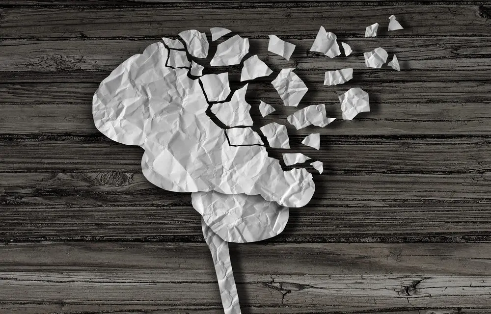
WHAT YOU NEED TO KNOW ABOUT TRAUMATIC BRAIN INJURIES (TBI)
Traumatic brain injuries (TBI) – or concussions – are a complex clinical phenomenon. The classic designations of mild, moderate, or severe TBI are based on the acute clinical presentation and do not necessarily predict the long-term outcome. Moreover, the long-held assumption that the mild forms of this condition recover rapidly and without consequence is not supported by more recent science. TBI leads to neurophysiological changes, changes in cell polarity, and premature cellular death. These consequences occur in sequence and can progress over a long period of time.
Mild TBI (mTBI), particularly repetitive mild TBI or concussions (as often happen in contact sports), can create tissue changes that lead to long-term increases in morbidity and mortality. As the extent of undiagnosed or undertreated mild TBI becomes more evident, the need to quickly identify any TBI, (particularly mild TBI) and then quickly provide effective treatments becomes increasingly important.
In 2003, the U.S. Centers for Disease Control and Prevention estimated the incidence of civilian TBI at 1.5 million. Globally, this number is estimated at closer to 10 million. Specific groups afflicted by TBI include an estimated 135,000 individuals per year from sports-related concussion alone and 82 per 100,000 of employees of the transportation industry. Meanwhile, the U. S. Department of Defense reported that more than 266,000 military Service Members experienced TBI between the years 2000–2012. The cost of TBI in the United States alone is considerable, estimated at over $76 billion per year in 2000. These estimates do not include the people who had bumps to the head without loss of consciousness. These individuals increase the numbers significantly.
In addition to the financial costs of TBI, the long-term decline in health of people with TBI is considerable. The rates of depression, anxiety, suicidality, drug and alcohol abuse, personality disorders, and other psychiatric symptoms are markedly elevated in those experiencing TBI. For example, elderly persons with a history of TBI have a higher risk for cognitive decline and potentially for Alzheimer’s disease than peers without a history. There is an additional increased risk of homelessness and higher rates of criminal behavior.
SYMPTOMS AND DIAGNOSIS OF TRAUMATIC BRAIN INJURIES
TBI is common in contact sports like football, hockey, and boxing. The short term consequences of acute brain injury are obvious – bleeding inside the skull or into the brain, and catastrophic brain injury – and may lead to death. But mTBI/concussion causes functional disturbance and injury to individual nerve fibers, meaning the injury is too subtle to be seen in most imaging studies, such as with an MRI.
Nerve cells, or neurons, are structured uniquely from other cell types. They are made up of the cell body itself, dendrites, and an axon. The cell body is similar to other cell types in its makeup. Dendrites are like the roots of a tree, spreading out from the cell body. Axons are slender, long projections that also come from the cell body. The axon of one neuron will generally connect with the dendrite from another neuron, creating a neural network that allows signals to transmit from one neuron to another.
When a concussion or TBI happens, it is the axon that takes the brunt of the damage. Axons are normally somewhat limber, but they become frail when exposed to rapid distortion such as happens with a brain injury. A frail or damaged axon can become inflamed. When normal communication from other neurons tries to take place with a damaged axon, even more damage occurs because the axon cannot handle the chemical or electrical information. It leads to a chain of events where multiple neurons are involved in the breakdown.
The extent of neuronal or axonal damage has to be significant to be seen on even the most sensitive imaging systems. Because of this, the diagnosis of TBI, particularly mTBI, remains a challenge clinically. There is no gold standard for diagnosis.
SHORT AND LONG TERM CONSEQUENCES OF TBIS
Following concussion, symptoms such as dizziness, nausea, reduced attention, amnesia and headache tend to develop acutely but usually resolve within a week or two. Severe concussion can also lead to loss of consciousness, which is a commonly-used criterion for assessing TBI in most neurological assessments. Despite the transient nature of the clinical symptoms, diagnostic assessments show that the disturbances seen with concussion take over a month to return to baseline. When tissue (neuro-pathological) evaluation is done, it shows that concussion-induced impairments may persist for years.
The clinical presentation can also be complicated by the overlap between the lingering symptoms of mild TBI and more long-term disorders, including posttraumatic stress disorder (PTSD), post-concussive syndrome (PCS), and chronic traumatic encephalopathy (CTE). These conditions can all exist separately or together. And, the symptoms of one can appear similar to the symptoms of another, making the diagnoses more challenging.
PERSISTENT SYMPTOMS FROM BRAIN INJURIES
About 15% of people who have suffered a TBI will develop lingering, persistent symptoms consistent with post-concussive syndrome (PCS). Depression and post-concussive syndrome (PCS) commonly present in a similar fashion. People suffering with PCS also report frequent headaches, dizziness, irritability and anxiety. To be formally diagnosed with PCS, a person must exhibit these symptoms for at least 3 months after the initial injury.
Chronic traumatic encephalopathy (CTE) is a neurodegenerative disease caused by repetitive mTBI, or “repetitive concussion”. People suffering from CTE used to be referred to as “punch-drunk” (since it was often found in boxers). CTE is characterized by protein deposits accumulating in neural tissues. At the moment, CTE can only be diagnosed pathologically, that is, by sampling a piece of the brain tissue, which is typically done after someone dies. Part of the reason it is so difficult to diagnose is because symptoms tend to only appear 8-10 years after the repetitive mTBIs themselves.
PCS is different than CTE in that the symptoms of PCS tend to resolve years before the onset of CTE. Proposed clinical research criteria for CTE include impairment in memory and executive function on neuropsychological testing. CTE should be considered in the differential diagnosis of a young adult with extensive repetitive head impact exposure and persistent mood and behavioral symptoms.
All of these conditions – a single TBI, repetitive mTBIs, PCS, and CTE – share common neuropsychological impairments, including memory loss, delayed problem solving, slowed reaction time, fatigue, and impulsivity. Such complexity can lead to misdirected treatment efforts and can hamper the ability to accurately assess treatment response.
Imaging using MRI is still not very helpful in identifying changes in brain function. By the time brain structure changes are seen by an MRI, the effectiveness of treatments may be more limited. Similarly, by the time pathological changes are observed, we have missed the opportunity to provide optimized treatment. As a consequence, structural changes may be insensitive to the earliest changes seen in the progression of damage in TBI.
It’s actually likely, given the limitations of every available diagnostic technology, that multiple approaches are necessary to provide a complete diagnostic picture.
CHANGES IN CELL FUNCTION (PATHOPHYSIOLOGY)
The development of CTE from a single TBI or multiple small, unrecognized mTBIs, is like removing bricks from the wall of a house: sometimes only one is removed at a time and sometimes larger sections are removed at once. The more bricks that are removed, the more obvious the damage is. By the time symptoms of CTE become obvious, many bricks have been removed permanently, lessening the ability to be able to create any impact on the condition or slow or reverse the progression of the condition.
Much in the same way that our bodies rely on our bones for structural support, individual cells need a cellular skeleton. One of the major components of the cellular skeleton is the microtubule. Microtubules are stabilized by proteins called tau proteins. Tau proteins are most abundant in neurons, although they do exist sparingly in other cell types. When a tau proteins become defective and stop stabilizing the microtubules, they lead to degenerative processes and diminish the ability of the cell axon to transport information. This leads to cellular death.
Focal axonal injury and spots of micro-bleeding lead to the deposition of abnormal tau proteins in the brain tissue – called the tau-positive neurofibrillary tangles (NFTs) – seen in dementia. All these changes point to the observation that acute TBI-related nerve cell injury, loss of microscopic nerve cell blood supply, breach of the blood brain barrier, resulting inflammatory cascade and activation of the defense mechanisms of the brain – microglia and astrocytes – are all likely to be the basis of the link between TBI and CTE.
The lesson from all this is that early, frequent, and continuing effective deep brain intervention is often necessary even with the earliest (even apparently innocuous) concussions/TBIs and minor head injuries. The brain is the most difficult organ in the body to recover structure and/or function in without outside stimulation and support from supplements and nutrition. The physiologic changes seen with concussion and TBI are the main targets for considering alternative therapies, such as pulsed electromagnetic field therapies (PEMFs).
TBI AND CONCUSSION RECOVERY WITH PEMFS
Because most current treatment approaches involve education, various individualized therapies, and symptom management, there is a significant need for consideration of other safe and non-toxic modalities to accelerate brain repair. Transcranial magnetic stimulation is one such potential therapeutic approach.
The science on PEMFs shows clearly that they penetrate all the tissues of the body equally, regardless of the tissue type. This includes the brain and nervous system. PEMF stimulation is applied externally, noninvasive, and simple to use. It may be low, medium or high intensity.
PEMFs applied to the head, with or without TBI, have been shown to have significant neural effects. PEMFs have effects on brain neuro-transmitter levels, affect monoamine function (such as dopamine, noradrenaline and serotonin), improve circulation and reaction time, increase stem cells and growth gene factors, and charge movement from neuronal membranes of cortical neurons. Even a very weak PEMF signal will stimulate about 25 billion neurons.
PEMFs have been proven to produce rapid mood elevation in depressed patients with bipolar disorder and other depressive disorders. Rapid transcranial magnetic stimulation (rTMS) is a robust, high intensity, FDA approved PEMF treatment for Major Depressive Disorder, and is also being studied in many neurological applications as a painless method to stimulate the brain. PEMF directed at the brain enhances neuroplasticity, entrains and resets brain cell resonance and communication between the thalamus, cortex and other brain regions, normalizes regulation and facilitates reemergence of natural cerebral rhythms, and through these mechanisms restores normal brain function. TMS can also be administered at a low magnetic field strength to affect multiple brain areas simultaneously.
RESEARCH ON PEMF FOR TBI AND CONCUSSION TREATMENT AND RECOVERY
Relative to TBI specifically, there are a number of studies using a range of different types of PEMF signals, from low intensity to very high intensity, with success.
One study explored whether PEMF signals could alter the course of inflammation in TBI. Inflammatory cytokine IL-1β production is increased in rats having experimental contusion or penetrating head injuries. IL-1β levels in cerebrospinal fluid (CSF) were proportional to injury severity in a contusion injury. PEMF treatment applied continuously reduced IL-1β levels by up to 10-fold in CSF within 6 hours after blunt injury and also significantly suppressed IL-1β within 17-24 hours after penetrating injury. This study clearly showed reduction of inflammation following head injury by a PEMF signal.
In another study, PEMF treatments as short as 30 minutes improved headaches following concussion. Patients with established diagnoses of mTBI with headache had average post-rTMS headache intensity reduced by 53%. The average headache exacerbation frequency (episodes per week) was reduced by 79%, with some patients reporting complete resolution of severe headache episodes.
Even a weak PEMF across the temporal lobes once per week for 5 weeks produced a significant improvement of depression and reduction of phobias in TBI patients.
rTMS has shown improvement with PTSD, pain, and integration between regions in the brain, along with improvement in related behavior and depression. One study found a 27% reduction in a depression score. rTMS can also improve cognitive function in Alzheimer’s Disease, a long-term consequence of TBI. rTMS improves brain activity through connected brain networks by improving cerebral blood flow (CBF) and therefore the supply of nutrients to brain nerve cells, not only at the stimulation site but, most importantly, in farther regions functionally connected with this site. Improving CBF can facilitate healing of brain tissues and improving brain function.
SAFETY AND RISK OF TREATMENT WITH PEMF
When PEMFs are suggested for the treatment of TBI, or for that matter, aimed at the brain for any reason, concern about safety and risk of brain harm is automatically raised. There is a great deal of evidence to suggest that there is minimal risk, with a large upside potential as seen from the literature review above. The safety and risk of PEMFs have been assessed in a number of studies.
Despite intense TMS or rTMS treatment programs, no significant side effects were seen. One patient received 70 treatment sessions over 12 months, or 420,000 pulses, with no side effects. One 75-year old patient received 130 sessions over 26 months with a total number of 156,000 stimuli, while 7 patients received 60 sessions over 12 months with a total number of 72,000 stimuli. In another study healthy men were given 12,960 high intensity rTMS magnetic pulses a day for up to 3 days in 1 week. This equals 38,880 magnetic pulses over 1 week, one of the largest exposures of rTMS to date. Despite this intense treatment regimen, no significant side effects were seen.
Even in the setting of relapsing remitting multiple sclerosis combined with TBI, no patient showed evidence of relapse during follow-up of at least 8 months. The authors concluded that magnetic brain stimulation was easy to perform, painless, and safe.
Some people also express a concern that EMFs might act as a cancer or seizure promoter. In fact, PEMFs appear to reduce the risk of seizures and do not promote brain glioma tumor growth.
Newborn brains are often considered to be especially vulnerable to PEMFs. High intensity magnetic fields applied even to newborn rat brains resulted in eight out of nine brain areas being thicker, suggesting improved brain cortex development.
HEALING CONCUSSION AND TRAUMATIC BRAIN INJURY WITH PEMF TREATMENTS
Traumatic brain injury and concussions have been an ever-present medical challenge for me as a doctor for over 40 years. The solutions to the problem today are little different than they were when I first learned about this problem in medical school. The biggest difference is that mild TBI, given the developing sophistication of medical knowledge, is now seen as a very important problem that needs to be dealt with sooner than later. In the past, only more serious brain injuries took our attention, typically those that involved admissions to intensive care for coma. Now we know that mild TBI, especially recurrent mild TBI’s leave very significant marks in the brain that result in major disability. The consequences of these TBIs have been brought to the forefront recently with the societal focus on sports concussions.
Given that most of the therapies for mild TBI’s are essentially adaptive, they help the body or person to cope or adapt to their disabilities, new approaches to managing this important condition are necessary. Modern evidence now suggests that even though somebody has recovered from their concussion, there are residual long-term effects in the brain. Other evidence indicates that the use of pulsed electromagnetic fields, of various kinds, early in the injury process helps to decrease one of the major aspects of the injury, which is inflammation in the brain.
The inflammation then causes all sorts of short-circuiting of brain function eventually leading to the symptoms, not only those seen in short-term, but also long-term: headaches, dizziness, depression, anxiety, insomnia, etc., etc. Additional evidence now also tells us that pulsed electromagnetic field therapies can help with, not only the injury itself, but also many of the symptoms resulting from it. In other words, PEMFs are not only useful for symptom management in the person suffering from TBI/concussion, but also have the opportunity to actually heal the brain to reverse the long-term effects of brain damage.
Medical management today, especially with medication, has limited value and is typically only useful for symptomatic management of the consequences of TBI, such as depression, headaches, memory issues, dizziness, etc. As such, medical management has almost no role in healing TBI other than facilitating adaptation and symptom management.
So, while there is evidence that pulsed magnetic fields, which reach deep into the brain and help all layers and areas of the brain without risk or side effects, there is a need for growing the medical knowledge base about the use of PEMFs for concussion/TBI, including establishing protocols for different PEMF systems for intensity, time and duration of treatment, frequency of treatment, and frequencies that are best used. It appears that even very high intensity PEMFs used for extended periods of time produce virtually no adverse effects on the brain and may even decrease the risk of future cancer development and the development of Alzheimer’s/dementia.
Meanwhile, PEMFs appear to have an important role in improving the damage caused by TBIs and the functional problems associated with TBI. Because of the lack of risk seen with the use of TBI, PEMFs should be a routine therapeutic approach in the management of mild to moderate TBI and concussion, and potentially greatly supportive to other adaptive therapies in accelerating recovery and function.