About 40% of people over age 65 experience some memory loss. It is known as “age-associated memory impairment,” if there is no medical condition that causes this loss of memory. Then, it is considered part of normal aging. Alzheimer’s disease and other dementias are not part of normal aging.
Age-associated memory changes are often seen as:
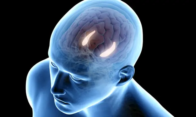
The hippocampus is responsible for memory and shrinks with age. It’s deep inside each brain, near the ear. It links unrelated things into memory, like where you left your keys or your neighbor’s name. It’s vital for forming new memories about experienced events. When it’s not normal, spatial orientation falters; people get lost, a common symptom of amnesia.
At Northwestern University, researchers studied PEMFs to stimulate older adults’ brains to improve memory. Since the hippocampus is deep, they targeted the parietal lobe, closely linked to it. Stimulation of the parietal lobe is expected to boost hippocampus function.
The study had 16 adults, ages 64-80. They compared memory tests with a younger control group before stimulation. Older individuals were correct about 40% compared to the younger group, around 55%.
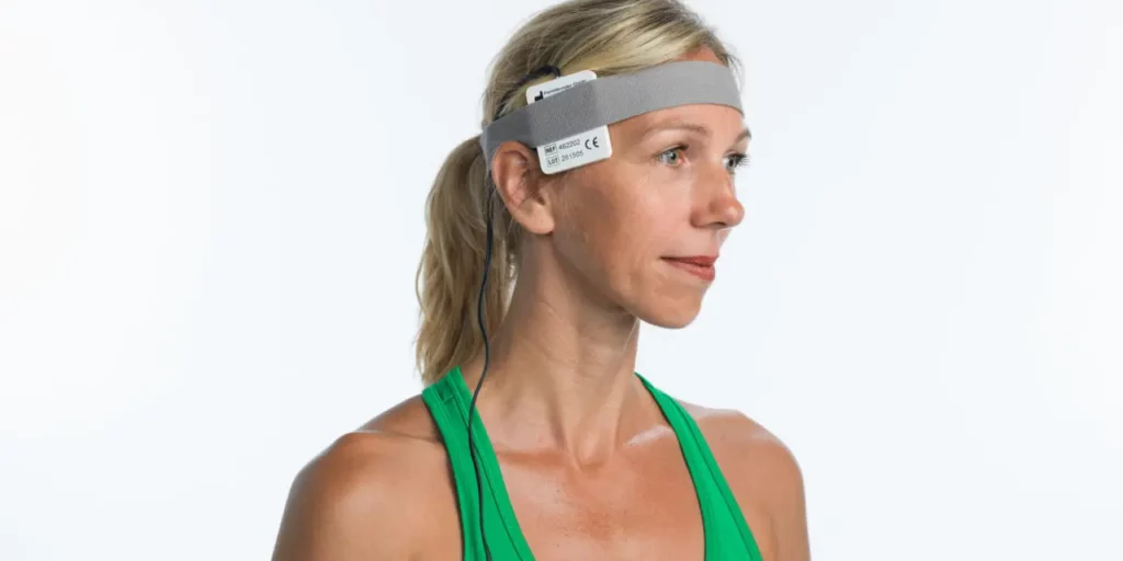
Stimulation was done using a high intensity PEMF at about 10 pulses per second for 20 minutes in each session, over five consecutive daily sessions to the left side of the head. Full intensity stimulation was compared to low intensity “sham” stimulation. This allowed the researchers to evaluate the changes in memory within the same individuals, a more realistic form of assessment, as would be seen in a clinical setting. At the end of the study. The researchers also used functional MRI (fMRI) of the brain to check brain function and the functional relationship of the parietal lobe with the hippocampus.
The results were as follows:
One of the lead authors said, “Older people’s memory got better up to the level that we could no longer tell them apart from younger people.”
This research also shows that stimulation of the parietal area of the brain impacts the memory associated with the hippocampus. The other words, the magnetic field penetrates deep enough into the brain through the parietal area into the hippocampal area to improve memory. The research also supports the idea that dysfunction of these connections increases with age, explaining the causes of memory loss as he will get older.
This research seems to indicate that the memory improvements do not last up to a week. It is not known whether longer episodes of stimulation would work better, whether stimulation beyond five sessions would work better, whether high intensity PEMF stimulation to other areas of the brain would produce similar or better results and whether stimulation with lower intensity PEMF systems would be as effective, and whether similar memory improvements could be seen in dementia or early-stage Alzheimer’s disease. Other high intensity brain stimulation research has already found benefits for Alzheimer’s disease. But, as is seen with many other conditions, the sooner treatment begins relative to memory loss the better the results. Waiting until Alzheimer’s disease has clearly been established is less likely to produce the same benefits as treating earlier age-related memory loss.
Nevertheless, these results are exciting, not only ensuring the safety of high intensity PEMF stimulation, but also that this PEMF therapy actually improves age-related memory decline.
Based on the results of this research I would recommend daily use of a home-based PEMF system, to encourage not only temporary improvement in memory but also to rebalance the tissues, hopefully age-reversing the age-related decreases in function of the hippocampal area. For convenience a portable PEMF unit may be likely to be used regularly, and certainly more affordable. Otherwise if someone already has a higher intensity PEMF system this can be used regularly to the parietal area of the brain, preferably daily.

Journal of Science and Medicine Abstract by William Pawluk discussing brain injuries
Read the full article in the Journal of Science and Medicine.
Traumatic brain injuries, or concussions, have challenged me as a doctor for over 40 years. Today’s solutions remain much as when I first learned about them in medical school. The key change is the growing recognition of mild TBI’s importance. In the past, serious brain injuries dominated attention, often requiring intensive care for coma patients. Now, we understand that even mild, especially recurrent cases, leave significant brain marks leading to major disability. Sports concussions have recently highlighted these consequences.
Therapies for mild to moderate TBI mainly focus on adaptation. New approaches are essential due to residual long-term brain effects, even after apparent recovery. Evidence suggests that early use of various pulsed electromagnetic fields can reduce brain inflammation, a major injury aspect. Inflammation disrupts brain function, causing both short-term and long-term symptoms like headaches, dizziness, depression, anxiety, and insomnia. These therapies can address the injury itself and many resulting symptoms, potentially healing the brain and reversing long-term damage.
Current medical management, often medication-based, mainly targets symptomatic relief for TBI consequences like depression, headaches, memory issues, and dizziness. It plays a limited role beyond symptom management and adaptation.
Evidence supports the use of pulsed magnetic fields, reaching deep into the brain without side effects. However, medical knowledge about PEMFs for concussion/TBI needs expansion, including establishing treatment protocols for different systems in terms of intensity, duration, frequency, and frequencies. High-intensity PEMFs used for extended periods appear to have no adverse brain effects and might even reduce cancer and Alzheimer’s/dementia risk.
Read the full article in the Journal of Science and Medicine.
Journal of Science and Medicine Abstract by William Pawluk about TBI
Read the full article in the Journal of Science and Medicine here…
This is a case report of a patient looking for a complementary approach to her chronic fatigue, and a mild partial complex, temporal lobe seizure disorder. Moreover, it is presumed the symptoms and seizure disorder were mostly attributed to her TBIs.
This report and discussion provide substantial support for the use of this specific 10 mT/100 Gauss pulsed magnetic field 10 Hz signal. This was for 2 hours daily to and only significantly improved clinical function. Also, it objectively produced positive neurological functional changes. Furthermore, it is still unknown whether 2 hours per day of this type of PEMF therapy is optimal. Clearly, in this patient, 2 hours/day of therapy made significant improvements in subjective and objective measures of function, and with internal validity, significant loss of benefit with cessation of therapy. Nonetheless, this loss of benefit with stopping treatment has been seen in other transcranial PEMF research. It remains to be seen whether durable, long-term benefits can be seen with longer-term PEMF therapy, whether other signal parameters could be optimized, including PEMF frequencies and intensities, and whether more permanent structural improvements in the injured brain may be found.
Further research is clearly needed on a larger sample of individuals with TBI. This is true whether complex or not, with different times after onset of injury. This would help to determine whether this approach is equally effective across various TBI scenarios. Along with this, whether any contraindications or limitations arise under different circumstances.
Read the full article in the Journal of Science and Medicine here…
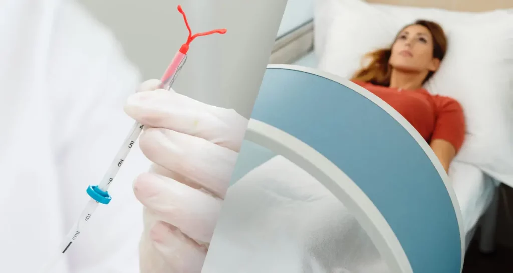
Long-acting reversible contraceptives (LARCs), such as intrauterine devices (IUDs) are in common use for pregnancy prevention. The percentages of use by age group are: ages 20–29 (13.7%), 30–39 (12.7%), 15–19 (5.8%) and 40–49 (6.6%).
Four IUDs are available in the United States, the copper-bearing IUD and three hormone releasing IUDs. Fewer than 1 woman out of 100 becomes pregnant in the first year of using IUDs (with typical use). IUDs are long-acting, can be removed, and can be used by women of all ages, regardless of history of pregnancy, and including adolescents.
A common question is whether using PEMFs in women with IUDs is safe. Three studies were found that looked at the question of the safety of doing MRI tests in the presence of IUDs. The only one that found any significant risk was for stainless steel -containing IUDs, which are not approved in the US.
Magnetic field interactions and potential adverse events were evaluated in 18 women using a questionnaire-based telephone survey. One woman reported a dislocation of the IUD after the MR test. All others had no signs of field interactions. The results were that MRI at 3.0-T can be used for women with copper-containing IUDs. (Berger-Kulemann) In another study, seven different types of copper IUDs were evaluated. Heating and dislocation of each IUD were investigated at two clinically relevant positions in 1.5 T and 3 T MR scanners. No significant heating of any tested IUD was detected during MR measurements. There was almost no temperature increase for all IUDs. No IUD movement was detected.
They concluded that there was no significant risk of possibly harming a woman was determined with an implanted copper IUD. (Neumann) the third study evaluated MRI safety of clinically used IUDs composed of copper/gold and stainless steel at 1.5T and 3.0T for displacement force, torque effects, and heating of IUDs composed of copper/gold (western IUDs) and stainless steel (China) on 1.5 and 3.0T MRI systems. They concluded that standard copper/gold IUDs can be considered as MR conditional for MR safety at 1.5 T and 3.0 T. The stainless steel IUDs, can be potentially harmful to during MRI due to high magnetic dislocation forces and torque. As a result, stainless steel IUDs (which are not available in the US or Canada) are considered MR unsafe. (Bussman)
Plastic IUDs would have no risk with PEMFs, since they do not contain metals. Drug-eluting IUDs have not been studied relative to MRI effects on the potential for enhanced drug release.
Therapeutic PEMFs, even high intensity systems, are not MRI machines. MRI devices contain both radiofrequency magnetic fields as well as high intensity static magnetic fields. High intensity radiofrequency are somewhat although minimally equivalent to pulsed magnetic fields. It can generally be assumed that even in women using high intensity PEMFs, that since they are MRI conditional, the safety/risk profiles can be considered acceptable. A major difference between MRI and PEMF treatment, is that PEMF treatment may be continued on a regular basis, whereas MRI is usually a relatively unusual, not frequently repeated. In this case we are particularly interested in the use of MRI in the pelvic area, and not other areas of the body.
Whether or not an MRI/high intensity PEMF is used, there is always the potential for dislocation of the IUD. Copper/gold metals are minimally magnetically attractable so there would be no torque on them to cause displacement. Very limited heating of the tissue when using a high intensity PEMF to the pelvic area is a possibility as the magnetic field bends around the metal, possibly somewhat increasing magnetic field intensity, in a very limited area around the IUD. The risk of this happening is the greatest with radiofrequency magnetic fields. However, these studies indicate this risk is almost nonexistent.
Another risk not discussed in the studies is the possibility of increased vaginal bleeding, due to the PEMF effect of decreasing platelet stickiness and increased fibrinolysis. Increased vaginal bleeding, especially around menses, is a natural consequence of an IUD in any event and may not necessarily be attributed to PEMF treatment. IUDs create additional risks of inflammation of the uterus and secondary infections in the fallopian tubes, ie, pelvic inflammatory disease (PID). PEMF stimulation may temporarily increase inflammation and potentially increase discomfort. However, PEMFs have been found useful in helping women with PID, along with antibiotic therapy.
If there is significant concern, especially with magnetic field intensities in excess of 7000 – 8000 Gauss, the duration of exposure to the PEMF may be reduced or the intensity kept to at least a 50% level.
Dislocation of an IUD is always a possibility even without MRI or PEMF exposure. However, if there is a concern or gynecologic issues develop, gynecologic examination would be recommended, particularly to see if there is dislocation.
There is almost no risk of using PEMFs in women with copper IUDs based on evidence from MRI studies. Usual risks associated wih the IUD are still present.