Heart Conditions
Table of Contents
ELECTROMAGNETIC FIELDS AND THE HEART: BASIC SCIENCE AND CLINICAL USE
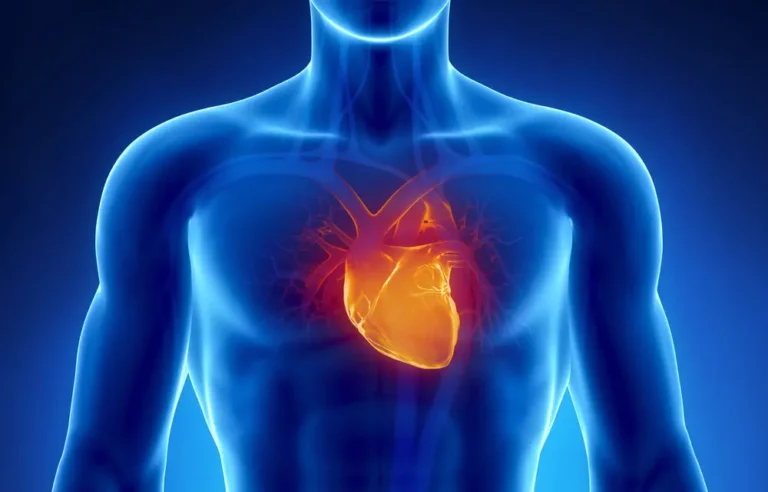
Heart disease is the number one cause of mortality in the United States and Canada. The heart is a very electrically dynamic organ. Heart disease includes many causes. These range from vascular disease, electrical conduction defects, muscle problems, valvular effects, congenital defects, infectious problems, trauma and pericardial problems, all as the direct or primary cause of the cardiac disease. Other non-direct problems can also affect the heart secondary to other systemic issues, including, but not limited to, hypertension, kidney disease, lung disease, autoimmune diseases, toxicities of various kinds, etc. Finding nonpharmacologic and noninvasive ways of managing heart disease in a safe, effective, nontoxic way is always a goal.
Magnetic fields have been found to significantly affect cardiac function, in addition to effects on a myriad of other body systems and problems. Not all magnetic fields are the same. Different types of magnetic fields may have different effects on the heart. Treatment of secondary causes can be just as important as the primary management of the heart itself.
The purpose of this paper is to explore the interaction of electromagnetic fields (EMFs) and the heart. We will review the basic science of EMFs and tissue, the heart as an electromagnetic organ and the geomagnetic and occupational influences that may affect it. This will serve to emphasize how readily cardiac tissue may be affected by EMFs. Animal studies often serve as a basis for understanding how human functioning may be affected by any therapy, before human treatment is tried. Finally, we will end with evidence of human benefits.
HOW DO MAGNETIC FIELDS AFFECT THE HEART?
EMFs either pass through the heart without interaction or they interact directly. The latter is called “coupling.” There are established, basic mechanisms through which static and time-varying electric and magnetic fields interact directly with living matter. Induced time-varying fields can stimulate excitable cells such as cardiac muscle. Static and time-varying fields interact with the body differently.
Every mammalian system reacts to the influence of EMFs. On the cellular level, cell membranes, mitochondria and nuclei are the most sensitive. Effects of EMFs depend on many factors, such as age (children and old people are more reactive), sex (men are more sensitive), and functional state (a functioning organ reacts more strongly).
NATURAL MAGNETIC FIELDS IN THE HEART
The heart muscle itself, because of its electrical activity, creates its own endogenous EMFs. Using a special magnetometer, one can see that the heart produces its own measurable, dynamic magnetic fields – electrical and magnetic activity follow each other, as night and day. These measurable fields allow for the mapping of the heart’s magnetic field under normal and pathological conditions and making possible a new tool for functional cardiology. EMFs generated by the cardiovascular system itself have biological effects not only on the heart itself but also on non-cardiac cells in the body distal to the heart by interacting with the immune system.
THE HEART AND ARTIFICIAL AND THERAPEUTIC MAGNETIC FIELDS
Extremely low frequency (ELF) EMFs easily penetrate tissues and cause virtually no sensory reactions. The reaction of the cardiovascular system to ELF EMFs is complex and includes direct responses of cardiac muscle, the autonomic nervous system, blood vessels, etc., and reflex responses mediated by the central nervous system. ELF EMFs increase the diameter of capillaries and greatly improve microcirculation, systemically and locally in the heart itself.
High frequency and high strength EMFs undoubtedly affect the cardiovascular system. Laboratory studies show that effects result even from EMFs below 150 Hz and 1G. The cell membrane is the primary site of EMF interaction, leading to intracellular changes in gene function and protein synthesis. These effects are highly nonlinear, with dose-response patterns that show “windows” of action and resonance-like phenomena. Power transmission lines can have health effects, but electrical transportation systems and electrical appliances are more common sources and have much more powerful EMFs.
Movement in the environment interacts with other external EMFs and work synergistically together to cause bioeffects. New lower limits to field strength actions are often discovered. There are no known lower limits for the intensity of EMFs to affect biological systems. Only the microscopic design of a receptor in the body and the time-variation dependency of its interaction with the many varied EMFs define the level of sensitivity to EMFs.
Extremely weak alternating, sinusoidal (power line) fields of certain frequencies interact with the local geomagnetic field and/or with EMF therapeutic systems. EMFs can act through another organ or tissue’s EM field, local and/or atmospheric geomagnetic fields and possibly even the moon. Even the body’s electrical currents interact with external fields and participate in control of life processes. External EMFs interact with most, if not all, organs and functions in the body. All these interactions clearly paint a very complex picture of EMF actions on the body – leading to the natural conclusion that human functioning is fundamentally inseparable from the EMFs around it.
ELF (below 300 Hz) EMFs interact strongly with biological systems, both electrically and magnetically. The magnetic field aspect of an ELF EMF penetrates the body without obstruction since the magnetic permeability of tissue equals that of air. Effects are directly related to the strength of the electrical currents induced in the tissue by the magnetic field aspect. Effects are seen at current strengths close to natural currents in the body and even fields well below those of natural tissues cause cellular responses. These involve membrane signal systems, cell surface receptors and all body biochemical systems, including enzyme activity and gene expression.
RESEARCH ON PEMF THERAPY FOR HEART CONDITIONS
In dogs’ hearts, considered comparable to human hearts, the natural heart EMFs, due to the beating of the heart, are much larger by adding external EMFs. The heart contributes between 5% and 10% of the total field induced in the human body by external electric and magnetic fields.
Cells or tissues can be protected against a lethal stress by first exposing them to a sublethal dose of the same or a different stressor to produce stress proteins in tissues. This concept is known as “preconditioning” and gives protection against oxidative stress, caused by ischemia/reperfusion, UV light exposure, heat, chemicals and electromagnetic field (EMF) exposure. Rodent heart muscle cells preconditioned by low energy EMFs for 30 minutes have more effective induction of stress proteins than heat. As little as 10 seconds of exposure produce a detectable response at 30 minutes, last for more than 3 hours and can be restimulated by a second exposure to fields of different intensity.
However, in egg embryo studies, continuous exposure to ELF EMFs for 4 days, twice daily for 30 minutes or 60 minutes for 4 days reduced the protective effect. 30-minute exposures once daily and 20-minute exposures twice daily did not reduce protection. A protective role is seen against cardiac ischemia in chick embryos. EMFs for 20 minutes induce stress proteins in the laboratory. This raises the strong possibility that using them before, during and after the surgical trauma can use EMFs for minimizing heart damage from heart surgery or transplantation or heart attack in humans.
For other kinds of cardiac actions, short-term exposure to sinusoidal ELF EMFs (5-8 Hz) in adult and old male rabbits for 15-120 minutes causes mild decreases in the ECG heart rate if exposure lasts 60 minutes. Old rabbits developed extra beats. In animals with experimentally induced myocardial infarctions, EMFs are not necessarily beneficial. There is little data comparing different kinds of EMFs. It remains to be determined what the optimal configurations are for different situations.
One study examined the difference between pulsed (PEMF) and alternating/sinusoidal (AMF) field effects on the hearts of dogs, exposed for an hour per day for 10 days. The AMF caused marked changes in heart dynamics: decreased ventricle function and increased peripheral resistance and end diastolic pressure in the right ventricle, as well as, left ventricular work. Systolic blood pressure (BP) and contractility and heart rate still decrease with PEMFs, but are less marked. Thus, use of PEMFs may be less aggressive for cardiac problems than sinusoidal fields.
Basic actions at the cell level account for these actions. A 16 Hz frequency modulation increases mean flow of Ca++ out of frog heart cells at low intensities. This compares with calcium flow in brain tissue, suggesting that neural tissues may generally react at these modulations and intensities and act through changes in Ca++ in and around cells. Chronic exposure to 50 Hz EMFs of rats at 2 hr/day for lower intensities or 0.5 hr/day to higher intensities produced increased blood flow to the heart tissue and enlargement of the coronary vessels. Higher intensity EMFs affect the heart function of rats (25) with 14 days, 4 hours a day of stimulation. EKG changes are temporary, but, at the end of 14 days of stimulation only heart rate remains decreased.
Hypertension, if untreated for a long time, can cause heart damage and ultimately heart failure. Static magnetic fields (SMF) of 2000 G placed on each carotid sinus area (south and north poles, respectively), decrease systolic, diastolic, and mean blood pressures by 10%. Heart rates were not affected. This action is most likely due to Ca++ transport changes across the carotid pressure receptor membranes.
Stress has very strong actions on the heart. When ultrahigh-frequency (UHF) EMFs are given to dogs subjected to emotional stress, several cardiovascular changes resulting from stress are improved. Stress increases blood pressure by 40-50%, heart rate by 20% and makes the left ventricle function hyperdynamically. Even though the UHF EMF does not eliminate the stress reaction of the cardiovascular system, it is less pronounced overall. UHF EMFs seem to accelerate central adaptation mechanisms, rebalancing circulation and decreasing adaptation time for cardiac stress.
HOW ENVIRONMENTAL AND GEOMAGNETIC FIELDS IMPACT THE HEART
Studies from Eastern Europe have found that changes in the geomagnetic field can worsen heart disease and that the major effects occur on the first or second day after a magnetic storm. Geomagnetic activity can be quiet, unsettled, active, or stormy.
While myocardial infarction rates (MIs) from circulation blockages do not seem to change with geomagnetic activity, cardiac electrical activity is probably more sensitive and affected. The admission rates of patients with new episodes of electrical conduction problems causing paroxysmal atrial fibrillation (PAF) were highest during the two lowest levels of geomagnetic activity, more in males and persons over 65 years. Males under age 65 with PAF are at greater risk of stroke from the PAF. Thus, increases in heart electrical instability appear to happen during periods of lowest geomagnetic activity.
Geomagnetic fields (GMF) also interact with ELF EMF therapy. Even a very weak EMF, up to 70 uT applied to the whole body or locally, for 8-12 minutes, 1-2 times per day for 10-20 days has clinical benefit in most patients. Sensitive patients improve after only one or two days, most others take 5-10 days. On days when GMF increases two-three fold, some patients complain of discomfort during the exposure and have increased blood pressure (BP). In most, (47%) the BP shows no changes, 38% decrease BP and 15% are increased.
Environmental exposure of EMFs also affects cardiovascular function. Average 50-60 Hz working environments do not have much effect on human heart rates. In AM radio station workers exposed to high frequency (HF) fields, 83% have heart rhythm disturbances and decreased signals in their ECGs.
There are significant differences between clinical ELF PEMF systems and high frequency (microwave or cell phone levels) sources. Any publicly oriented article or book, unselectively citing references mixed with ELF exposures and those in the HF kHz and over range, clearly does not understand EMFs and is indiscriminately comparing apples and oranges, in terms of their clinical effects. Indeed, there are many clinical therapy systems that use high frequencies, but they are usually used for tissue destruction, for tumors and colon, bladder, skin and heart arrhythmia lesions, etc. General or public use of EMFs for personal use should be restricted to low strength ELFs or high frequency EMFs that do not create heating.
Duration of exposure to environmental fields is probably important as well. Changes in heart rhythm may be affected in the workers professionally exposed to 50-Hz electric and magnetic fields (EMF) over long periods. There can be a global decrease of cardiac rhythm in both high (over the industry norm) and low (at or below industry norms) professionally EMF-exposed groups compared to the non EMF-exposed control group. These changes may increase the risk of cardiovascular diseases. Other environmental or home-based “electromagnetic pollution” has the risk of inducing health problems. Fortunately, measures can be taken in the home and office to decrease background EMF risks.
HUMAN STUDIES ON PEMF THERAPY FOR HEART CONDITIONS
Experimental studies show that EMFs can affect the function of the centers of the autonomic nervous system controlling cardiac rhythm. A temporary increase in BP is seen with clinical exposure to industrial 50-60 Hz EMFs, but extended exposure causes the systemic pressure to decrease. Microcirculation dilatation occurs, with increased blood flow in the capillary bed and precapillary arterioles and an increased permeability of the vascular wall. Even lymphatic vessel flow increases. Circulation changes produced by EMFs are depending on the functional state of the central regulatory apparatus, especially the hypothalamus. Experimental PEMFs are found to act directly on the tissue of a beating heart.
Medium powerline-type field exposure for 3 hours causes a significant slowing of the heart rate. EMF effects are related to changes occurring during the recovery phase of the cardiac cycle. Humans are more responsive to some combinations or levels of field strength than others.
EMF therapy acts beneficially on the functional state of the nervous and endocrine systems as well as on tissue metabolism. The heart rate and BP decrease and the cardiovascular system is less reactive to adrenaline and acetylcholine. The parasympathetic nervous system is activated. Stimulation of the autonomic ganglia along the spine reduces cortisol and aldosterone. MFs typically cause only a momentary change of the microvascular bed with slowing blood flow. This then changes over to a longer period of an increased heart rate, rate of blood flow and filling of the blood vessels.
HOW PEMF THERAPY BENEFITS THE HUMAN HEART
Sinusoidal PEMFs improve microcirculation in people with ischemic heart disease and vascular diseases of extremities. PEMFs act more strongly than permanent magnets. ELF MFs improve both lipoproteins and cholesterol levels. But, a static magnetic field of more than 50 mT (500 Gauss) at the tissue increases risk of atherosclerosis, with irregularly arranged lipid deposits in middle to large size arteries and fibrosis and calcification. In people with low blood pressure, EMFs improve heart contractions and cause more normal bioelectrical function. In most people, EMFs lower BP by lowering vascular resistance, with vasodilatation.
Hypertensive patients are affected positively, depending on the function of the heart before magnetic treatment. People with normal functioning hearts just have their vascular resistance lowered. EMFs normalize heart function and circulation in patients with high BP, and at the same improve circulation. The improvements in systemic vascular tone, as well as lipid metabolism and coronary circulation make MFs very useful treatment for people with the combination of hypertension and ischemic heart disease.
Early in the course of use of MFs in patients, there are changes in ECGs to a lower wave size pattern, sinus rhythm and extra beats, and a decrease in heart rate. With continuing magnetotherapy, these changes disappear and cardiovascular function is improved. This is common with MF therapy. Meanwhile, there may be temporary worsening while repair and rebalancing is happening, with the outcome being more normal function and health.
To get better results with EMF treatments, understanding the underlying cause of the problem and function of the organ system is critical for designing the proper protocol to use for an individualized approach. The best outcomes occur this way. Without an understanding of the physiology and the type of field to use and how, less than optimal results can happen. Awareness of the potential for initial de-stabilization minimizes misunderstanding in managing the course of therapy and should be carried out with the assistance of a knowledgeable professional.
MAGNETIC THERAPIES TO AVOID FOR HEART CONDITIONS
Good results are not always seen. In one small series, patients were treated with sinusoidal EMFs for arrhythmias caused by ischemic heart disease, post myocardial infarction and cardiomyopathy. A sinusoidal EMF used for 10 sessions daily, alternating between placement to the sternum for 15 minutes and “palm – wrist” area for 5-7 minutes. EMFs did not normalize heart rhythm. One woman had an attack of paroxysmal tachycardia occurred. Six patients reported unpleasant sensations (“sickness at heart” and headache) during or after EMF therapy, occurring most often with cardiomyopathy. A sinusoidal EMF may even increase BP in males, whether exposure was for 20-40 minutes or 1 hour.
OTHER MAGNETIC FIELDS AND THE HEART
Magnetolaser therapy (MLT) has been studied in single placebo control trial in the treatment of ischemic heart disease patients, with exertional angina and moderately to severely impaired function, post-myocardial infarction. Most had significantly decreased circulation. MLT was applied to 3 tender zones on the chest: in the front over the upper part of the heart and middle of the sternum, and in back between the scapulas to the left of the mid line, for 12 min, 4 min for each exposure zone, daily over 15 days. Work capacity increased in 84% of the MLT group but worsened in the placebo group.
The work increased most for patients with functional classes II and III angina. MLT was also useful for patients with conduction disorders, eliminating extra beats in 29% and decreasing them by more than 70% in 32% of cases and stopping paroxysmal atrial fibrillation in 53%. The treatment lasted through the follow-up period of 12 to 16 months. These impressive results show that MLT facilitates adaptation to a physical load, and promotes rearrangement of central hemodynamics and recovery and stabilization of electrical activity of heart cells, safely and simply.
Heart rate variability (HRV) results from the action of neuronal and cardiovascular reflexes, including those involved in the control of temperature, blood pressure and respiration. Changes in HRV are predictive of a number of cardiovascular disease conditions and specific alterations in HRV have been widely reported to be associated with adverse cardiovascular health outcomes. Low strength, 60-Hz continuous or intermittent MFs in healthy males has little or no effect on HRV, indicating they do not induce stress effects. HRV alterations during magnetic field exposure may occur when accompanied by increases in physiologic arousal, stress, or a disturbance in sleep. There appear to be significant differences in heart rate and mean 24-hour personal exposure to MF between occupational and non-occupational group. It is not yet known whether clinical EMF exposure, in those who’s HRVs shows clear departures from normal, improves the HRV.
Patients with so-called Electrical Hypersensitivity (EHS) have a misbalance of autonomic regulation being more hypersympathetic, as measured by heart rate (HR) and electrodermal activity and sympathetic skin responses to visual and audio stimulation. There are frequency-intensity-duration components to these exposure sensitivities.
Much of the biological effect of high frequency fields is due to tissue heating, not just EMF effects. However, biological effects, not due to tissue heating (nonthermal) have been found with millimeter-range (MMR; about 300 GHz) MFs. These EMFs probably affect the command centers of organs through reflex systems. It is preferable to select biologically active points for MMR exposure; tender zones and areas of large joints, where tissue sensors are numerous and nerve fibers contact collagen directly. Local skin exposure to MMR EMF affects cerebral function.
Clinical benefits are seen with treating heart angina and hypertension, especially essential hypertension. It is noted that patients with symptomatic, renal hypertension don’t respond. Cardiac rehabilitation in rats with MMR EMF added after myocardial infarction promotes tissue repair and functional recovery. Human clinical studies confirm this. Only favorable effects occur: more rapid tissue healing, activation of ATPases, antioxidant properties, and so on.
MOBILE PHONE EMISSIONS AND THE HEART
Use of mobile phones is commonplace. Mobile phones produce biologically active HF EMFs. Digital mobile telephones have the theoretical possibility of affecting implanted cardiac pacemakers. The META series of pacemakers are mostly immune to clinically important EMF interference from digital mobile telephones. It appears that use of digital mobile telephones by patients with appropriately programmed pacemakers is safe.
There is also much evidence of accelerated tissue healing effects where there has already been tissue damage, either from ischemia or mechanical or surgical trauma. Tissue healing with PEMFs appears to be accelerated by about one third to one half the usual time and results in fewer tissue complications, such as infections and poor or aggressive scar formation. In addition to stress protein effects mentioned above, but also free radical scavenging and accelerating RNA/DNA production in laying down new repair tissue. This means that, if cardiac surgery is required, not only would EMFs help tissues to be less traumatized but also would help speed recovery afterwards.
Cardiac vascular and peripheral vascular blockages depend on the development of “soft” and “hard” atheromatous plaque formation. It is these plaques and their actions on platelet dynamics that can cause vascular obstructions, occlusions or thromboses, leading to cardiac ischemia (angina) or heart attacks. This is the basis for several drug therapies aimed at reducing platelet adhesiveness, including daily aspirin and warfarin, to name a few. PEMFs have similar, strong effects in reducing platelet function and other clotting factors and probably acts synergistically and additively with these drugs. The primary actions studied have been on ADP dependant platelet adhesion, reduction in thrombin and increased thrombolysis.
MAGNETIC THERAPY AND THE HEART – WHAT YOU NEED TO KNOW
EMF therapy acts beneficially on the functional state of the cardiovascular, nervous and endocrine systems as well as on tissue metabolism. The heart has been found through numerous studies to be very electromagnetically sensitive. This sensitivity extends to all external fields, whether therapeutic or otherwise, including their interactions with each other and with the body.
Some of the benefits to the cardiovascular system are indirect, acting through stress reduction effects, emotional responses, endocrine system, the immune system and especially the autonomic nervous system. Autonomic neural regulation makes the tone of the vascular system normal. Even cardiac muscle blood vessels are dilated. Decreased vascular resistance decreases the workload of the heart, reducing strain, which if applied over long periods of time could lead to decreased cardiac wear and tear. EMFs have a moderating effect on cardiac function as well as the microcirculatory system. Pulsed magnetic fields, versus sinusoidal fields, appear to be less aggressive towards the heart. Much of their actions depend on cellular Ca++ ion changes.
There are multiple other actions of EMFs on the cardiovascular system. One is an anti-atherogenic effect and a reduction of platelet adhesion factors, which could reduce the possibility of cardiac vascular occlusions and cardiac damage. Because of actions on stress proteins, cardio-protection is now a feasible use, not only for treating or reducing cardiac ischemia but also for the trauma created by cardiac surgery. EMFs are even useful post-operatively in facilitating and accelerating recovery through wound healing effects, for superficial and even deep tissues.
There are significant differences between clinical ELF PEMF systems and high frequency (microwave or cell phone levels) sources. Any publicly oriented article or book, unselectively citing references mixed with ELF exposures and those in the HF kHz and over range, clearly does not understand EMFs and is indiscriminately comparing apples and oranges, in terms of their clinical effects. Indeed, there are many clinical therapy systems that use high frequencies, but they are usually used for tissue destruction, for tumors and colon, bladder, skin and heart arrhythmia lesions, etc. General or public use of EMFs for personal use should be limited to low strength ELFs or high frequency EMFs that do not create heating.
The evidence reviewed here gives reasonable support for wider medical application of magnetic field (MF) therapy as a method of non-drug therapy in cardiovascular disease, alone or in a complementary fashion with medical or other modalities.
